


Project Description
Our group has shown left-lateralized reductions in MR volume of the amygdala-hippocampal complex and parahippocampal gyrus, regions important to verbal memory and associations, and areas that are highly intercorrelated with each other and with the superior temporal gyrus (see the “Projects” section entitled “Chronic Schizophrenia”). These findings suggest that damage to an interconnected neural network in the brain may reflect damage that is functionally important for associative links, and may lead to difficulties in the storage of auditory and/or language information. To investigate this further, our group will be looking more closely at language areas in the brain.
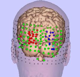
A three-dimensional model of the brain and skin, with areas of TMS activation shown in red and blue.
The goal of our TMS project is to use a new investigative tool, transcranial magnetic stimulation (TMS), to map cortical motor and speech areas in the human brain. As a procedure, TMS involves the use of a current pulse that passes through an insulated coil held over the subject’s head. The current pulse induces a pulsed magnetic field, which like other forms of electromagnetic waves, passes through the scalp and skull without attenuation. This current pulse causes the excitation of cortical neurons. If the current is in motor cortex, peripheral myographic responses are observed (i.e., movement of the finger). If it is in cortical speech areas, a disruption and/or inhibition of verbal processes are observed. TMS thus has the potential for a broad application in neuroscience because it can be used to evaluate more closely the relation between brain structure and function in both controls and disorders characterized by cognitive impairments such as schizophrenia.
Our study focused on the co-registration of MR with diodes on the skin using a pixel camera system, and diodes placed on the TMS probe in order to co-register coordinates in MR with TMS, and skin (Ettinger et al. 1997). A further study has evaluated motor cortex maps using TMS (below). In another study we inhibited visual perception using occipital TMS (Potts et al., 1998). This visual suppression effect has previously been attributed to the disruption of gray matter of primary visual cortex or in the fiber tracts leading to V1 from thalamus. However, others have suggested that this visual suppression effect may be due to a disruption of secondary visual cortex. In this study we produced visual suppression contralateral to the stimulated hemisphere in five normal volunteer subjects (Potts et al., 1998). We coregistered the stimulation sites with magnetic resonance images in these same subjects using optical digitization. The stimulation sites were mapped onto the surface of the occipital lobes in three-dimensional reconstructions of the cortical surface in order to show the distribution of the visual suppression effect. The results were quite consistent with disruption of secondary visual cortical areas.
Work currently involves mapping motor and language areas in both controls and in patients prior to neurosurgery. Our goal using neurosurgery patients is to determine how closely functional regions mapped by TMS correlate with direct brain stimulation intraoperatively.

Comments are closed.