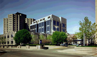
The main mission of this laboratory is to understand brain abnormalities and their role in neuropsychiatric disorders using state-of-the-art neuroimaging techniques. We began our work in neuroimaging studies in schizophrenia in the late 1980’s in the Surgical Planning Laboratory (SPL), MRI Division, Department of Radiology, Brigham and Women’s Hospital. This research collaboration grew out of early conversations between Drs. Robert McCarley, Director of the Neuroscience Laboratory, and Professor and Chair of Psychiatry, Harvard Medical School at Brockton VAMC, and Ferenc Jolesz, Holman Professor of Radiology and Head of the MRI Division, Brigham and Women’s Hospital, Harvard Medical School. This research moved from CT studies in 1986 to MRI, with the latter studies put on solid ground in the late 1980’s with a Research Scientist Development Award (K01) from the National Institute of Mental Health (NIMH) to Dr. Martha Shenton, and Dr. Ron Kikinis’ arrival to the MRI Division in Radiology.
The Psychiatry Neuroimaging Laboratory (PNL), Departments of Psychiatry and Radiology, Brigham and Women’s Hospital, Harvard Medical School, was founded on October 1st 2005, with Dr. Martha Shenton as Director. In 2008, Dr. Marek Kubicki and Dr. Sylvain Bouix became Associate Directors. The research conducted in the PNL has expanded to include a number of junior faculty, post-doctoral fellows, graduate and undergraduates, from multiple disciplines, who are investigating the role of brain abnormalities in not only schizophrenia but also post-traumatic stress disorder (PTSD), traumatic brain injury (TBI), Attention-deficit hyperactivity disorder (ADHD), velocardiofacial syndrome, and William’s syndrome. It is well funded through grant support from private foundations, NIMH, NINDS, VA Merit Awards, and more recently, from the Department of Defense, where Dr. Shenton and colleagues are involved in a large Clinical Consortium, INTRuST, to understand PTSD and TBI, with an number of researchers including Dr. Ross Zafonte. Work has also begun in sports-related head injuries. Here, the Department of Defense is funding a positron emission tomography (PET) study to investigate the role of tau pathology in living retired National Football League players, in collaboration with Drs. Robert Stern, Alex Lin, Inga Koerte, as well as others. A research project has also begun to investigate subconcussive blows to the head in elite professional soccer players in collaboration with Drs. Inga Koerte, Birgit Ertl-Wagner, Maximilian Reiser, Ross Zafonte, and Robert Stern. The impact of head trauma is also being investigated in university hockey players with Drs. Paul Echlin, Inga Koerte, Ofer Pasternak, Karl Helmer and Sylvain Bouix. All of these advanced neuroscience studies utilize tools and technologies developed in the PNL, which include (but are not limited to): 1) a multi-tensor tractography algorithm (developed by Dr. Yogesh Rathi) to trace white matter pathways in the brain, 2) a free-water imaging technique (developed by Dr. Ofer Pasternak), which provides information about the structure of the tissue, 3) an algorithim to measure the geometry of white matter fibers (developed by Dr. Peter Savadjiev), and 4) a compressed sensing based algorithm for faster acquisition of diffusion MRI scans (Dr. Yogesh Rathi). Without such advances in imaging technology the neuroscience studies would not be possible.


Comments are closed.