


Project Description
The quantitative analysis of magnetic resonance imaging (MRI) data to examine anatomical brain structures is a fundamental component in the assessment of structural brain abnormalities. The method currently used for analyzing brain structures involves a laborious manual tracing of the contours of anatomical structures derived from MRI scans. The time requirements for this particular method make it practically impossible for a research team to measure more than a limited number of brain structures in a patient population and in a matched control group. An automated procedure would thus greatly increase both the number of regions as well as the number of individuals who could be investigated in any one study.
An automated technique for quantitative MRI analysis would have to be able to differentiate between structures with similar intensity on the MRI scans. One solution would be to use an anatomical atlas as a template, in order to provide anatomical information not obvious from MR contrast alone. We have used such an approach in our work, where we have used an MR atlas as the template for elastic matching of these same structures in new MR brain scans.
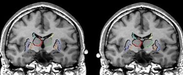
Measurements of the basal gangia done manually (left) and using elastic matching techniques (right).
In one study, we used an MR brain atlas, developed in our laboratory in order to evaluate, and compare, our automated registration algorithm for the volumetric measurement of MRI brain structures, to the same measurements performed using manual tracing. Accordingly, we took information from the MR brain atlas, based on one control subject, and projected it into other MRI scans by applying an elastic match (i.e., warping the atlas into the shape of the new brain image). In this study we were able to measure, with high accuracy, the volumes of eleven brain structures using elastic matching. (These brain structures included: total brain volume, total gray matter, total white matter, left and right thalamus, left and right caudate, left and right putamen, and left and right globus pallidus; see Iosifescu et al., 1997).
Elastic matching can increase the possibility of highlighting subtle morphological changes associated with schizophrenia and other chronic brain disease. Hence, our research team continues to work with computer scientists and other researchers at the SPL in order to develop new and improved tools for making automated measurements. At the same time, continuously updating and improving our brain atlas remains a priority.

Comments are closed.