Project Description
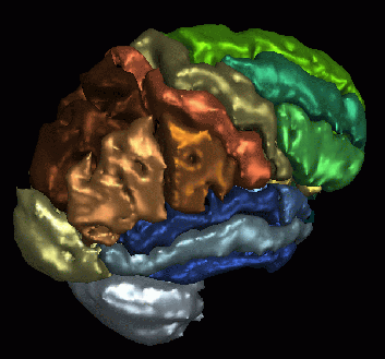
Learning neuroanatomy can be difficult because of the tremendous amount of detail and complexity in the interrelationships among neuroanatomical structures. The use of three-dimensional (3D) anatomic models (or atlases) can greatly facilitate the learning of anatomy because they provide the student with important information about not only the shape of a structure but also its spatial relationship to surrounding structures. Other important features of 3D graphical models are the ability to manipulate the models by removing or making transparent various structures, and the ability to zoom in to examine the detail of structures.

Our brain atlas allows for interactive manipulation of 3D models of the human brain on personal computers without high transmission costs or the need for specialized graphics hardware. The main features of the browser include the ability to display various views of the model, to select structures to be annotated, to remove or make structures transparent, to zoom in on structures, and to collapse and expand a hierarchical description of structures to control the level of detail displayed. An additional goal in creating the brain atlas is to use it as a template for automatically segmenting regions of interest in new MR data sets. To date, our atlas has more than 150 neuroanatomical regions delineated. These brain regions can be identified in new MR data sets using coregistration and elastic matching (or warping) techniques. The latter is described in more detail in the section entitled “Elastic Matching Studies”. Our work with the brain atlas has now been extended to other parts of the human body, including the chest and abdomen, spine, pelvis, inner ear and knee, as well as to other techniques.
We have also been investigating the creation of a quantitative atlas, which can capture the distribution of diffusion MRI measures within a normal population. This normative atlas has been used to detect abnormal measurements in subjects suffering from persistent post-concussive symptoms which were not detected by neuroradiologists (Bouix, Pasternak et al., 2013).


Comments are closed.