


Lab News

Brigham Researchers Awarded Two Prestigious NIH Grants
December 8, 2020
Textbook on the use of MRI in psychiatry
September 2, 2020General News
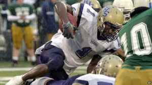
NCAA offers settlement in athlete concussion cases
July 31, 2014Message from the PNL Director
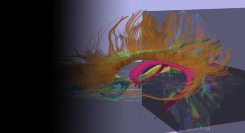
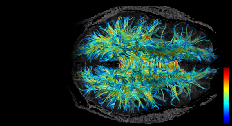
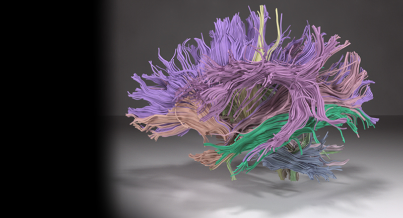
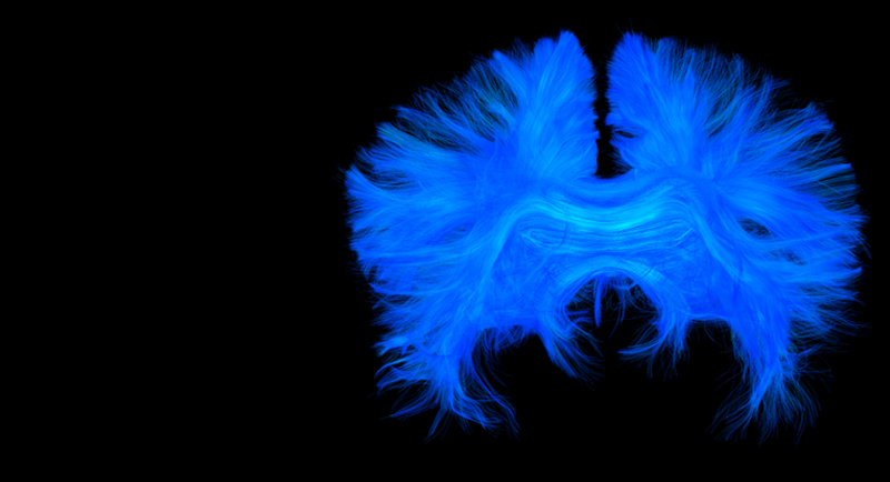
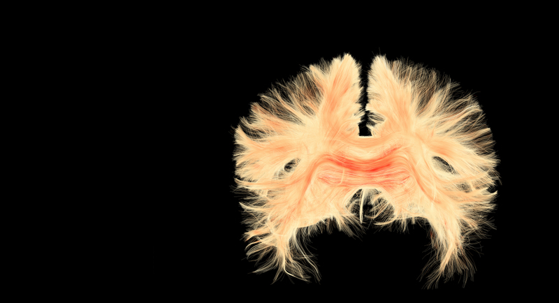
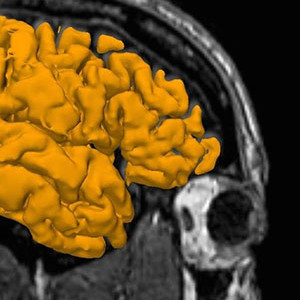
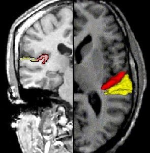
Visualizing White Matter
Cortical Spinal Tract (Yellow) and
Cingulum Bundle (Red)
Research Area
Education

NPR: US Science Suffering From Booms And Busts in Funding
September 9, 2014
NPR: When Scientists Give Up
September 9, 2014People
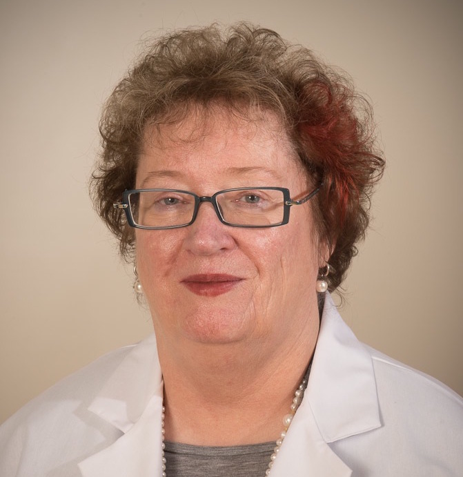
Martha E. Shenton, Ph.D.
July 9, 2014
Marek Kubicki, M.D., Ph.D.
July 8, 2014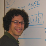
Sylvain Bouix, Ph.D.
July 8, 2014

Comments are closed.