Project Description
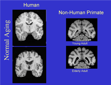
The ability to identify and follow structural brain changes during brain maturation and aging is both fundamental to our understanding of brain function, and crucial in clinical studies. While post-mortem studies of human brain can provide data on local histological changes, they are only cross sectional. Further, such brain samples are not optimal and the sample sizes are generally small. In contrast, non-invasive in vivo imaging can provide powerful longitudinal data for large populations. Moreover, post-processing techniques make it possible to analyze the entire brain, providing anatomically specific data that allows for investigating relationships between imaging and function. Recent developments in diffusion MRI and fiber tractography have revealed correlations between imaging changes and cognitive aging in both monkeys (Makris et al., 2007), and humans (Voineskos et al., 2012). Unfortunately, the biological  underpinnings of such imaging changes are largely speculative (Paus 2010) and hence the specificity of imaging measures for histological features is unknown. The lack of such “validation” is largely due to the inability to conduct well-controlled studies of both brain tissue and imaging in humans. We are conducting an NIH (NIA and NIMH) funded (1R01 AG04252; Neural substrates of diffusion imaging in cognitively aging rhesus monkeys) multidisciplinary study using a rhesus monkey model of normal aging. This is enabled by a collaboration of three labs (Animal Cognitive Neurobiology Lab at BU, Center for Morphometric Analysis at MGH and the PNL), with unique and complementary expertise in MRI imaging, morphometry, neuroanatomy and cognitive aging. We have available a cohort of over 50 normal aging rhesus monkeys of both sexes, ranging in age from 5 (young adults) to over 30 (oldest of the old) years of age. Most important is the availability of cognitive and DTI data that can be used for histopathological validation of archived, cryoprotected, unstained tissue from all of these monkeys. The main aims of this study are: 1) to establish sensitivity of individual diffusion tensor imaging (DTI) metrics to cognitive maturation and aging; 2) to investigate biological underpinnings of diffusion changes during cognitive maturation and aging; and 3) to develop free imaging tools necessary to accomplish the goals of this proposal. The results of this study will greatly impact our understanding of aging processes and their mechanisms and provide tissue validated understanding of imaging measures that can be applied to studies of normal human aging as well as many other clinical populations including traumatic brain injury, multiple sclerosis, and schizophrenia.


Comments are closed.