


Project Description
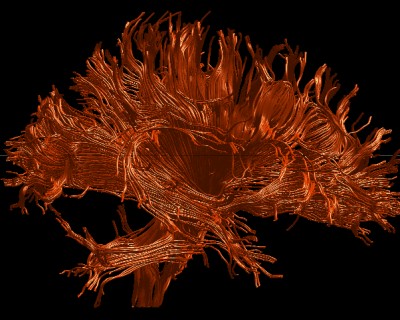
Diffusion Tensor Imaging (DTI) is a relatively new imaging technique that can be used to evaluate white matter in the brain. Using DTI, the orientation and direction of white matter fiber tracts can be visualized and quantified (see Figure below which shows visualization of white matter fiber bundles in the brain). This method involves quantifying the orientation and directional uniformity of water diffusion in brain tissue. In white matter, the mobility of water is restricted in directions perpendicular to the axons oriented along the fiber tracts, and thus the orientation and direction of these fibers can be traced. We plan to use these techniques to evaluate the orientation and asymmetry of white matter fiber tracts in patients diagnosed with schizophrenia (chronic, first episode, and those at high risk for schizophrenia – prodromes), as well as in patients diagnosed with affective psychosis (e.g., bipolar disorder), patients diagnosed with velocardiofacial syndrome (a chromosomal deletion on chromosome 22 that has a high risk for schizophrenia and psychosis), patients diagnosed with William’s syndrome, patients who are suffering from traumatic brain injury, and in control subjects (as measured by geometric measures of diffusion including, linear, planar and spherical diffusion).
We predict that connections between the hemispheres will be disrupted in schizophrenia and that these connections will be more disrupted in the left hemisphere in patients with more positive symptoms, but bilaterally in patients with more negative symptoms. We further predict that these abnormalities will be most evident in links between frontal and temporal lobes. Accordingly, in one study we have shown diffusion abnormalities in the uncinate fasciculus, the largest fiber tract connecting the frontal and temporal lobe, in patients diagnosed with chronic schizophrenia compared with normal controls (Kubicki et al. 2002; see also review by Kubicki et al 2007). We plan to also follow first episode patients where we predict that the percent change over time in diffusion abnormalities will be greatest in first episode patients with schizophrenia compared with affective patients and controls. We also predict that connections between the hemispheres via the corpus callosum will be more dense in schizophrenia and show an increase in diffusion, which would be consistent with the long-held theory of too much communication between the hemispheres. We will apply these same techniques to understand further white matter abnormalities in velocardiofacial syndrome, William’s syndrome, traumatic brain disorder, and in animal models including the aging brain in rhesus monkeys (see Research Area and publications for specific details).

Comments are closed.